
DOWNLOAD THE APP


Customer Support

Desert Online General Trading LLC
Warehouse # 7, 4th Street, Umm Ramool, Dubai, 30183, Dubai
DOWNLOAD THE APP
Customer Support
Desert Online General Trading LLC
Warehouse # 7, 4th Street, Umm Ramool, Dubai, 30183, Dubai
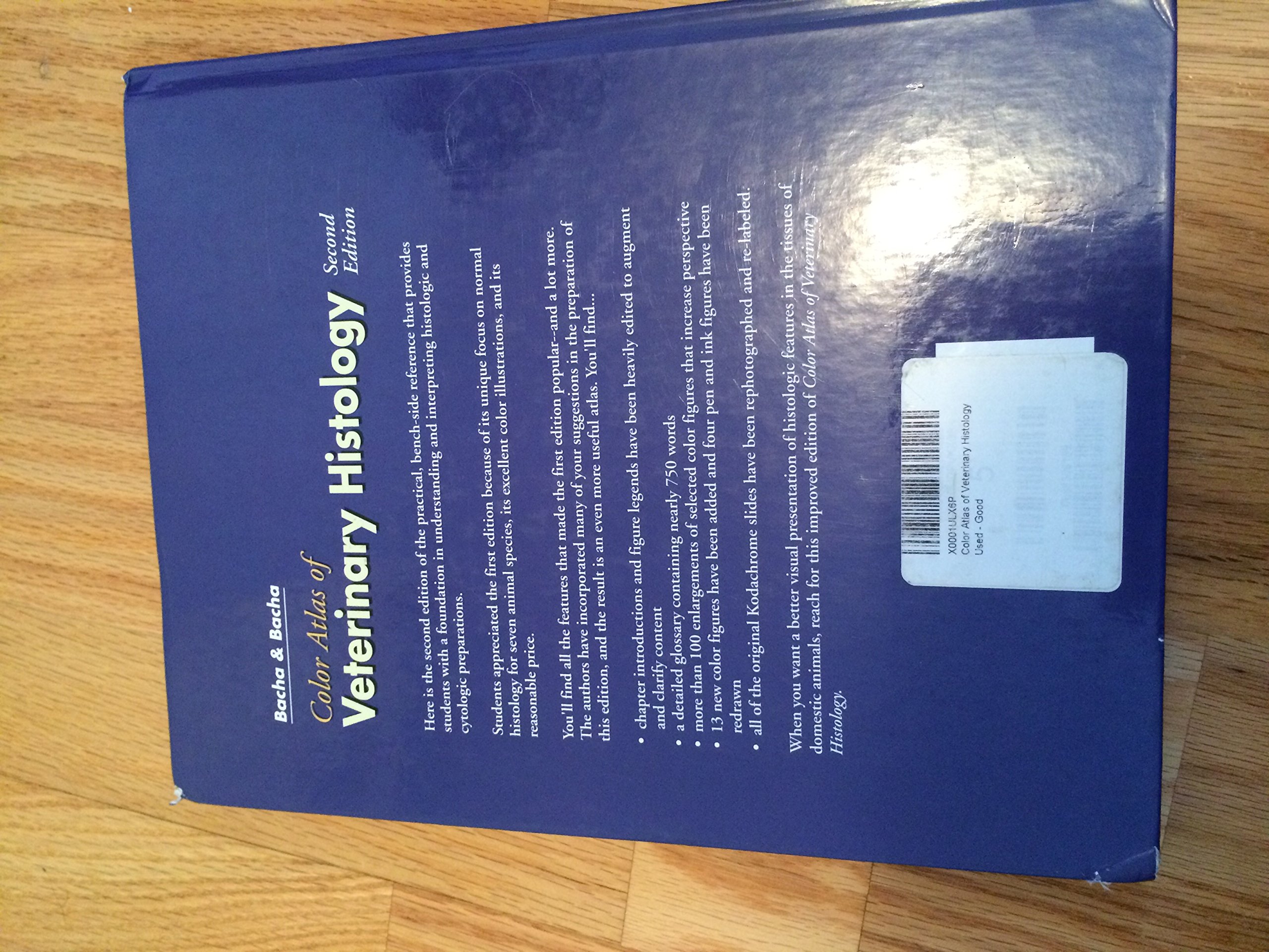
Full description not available
C**E
Five Stars
Perfect for histology my first year. I will definitely pass this on when I graduate.
C**B
Five Stars
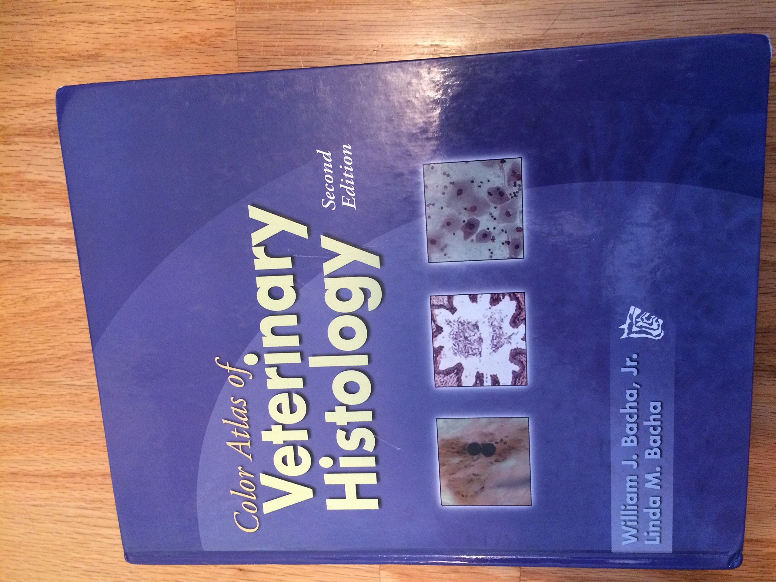
Excellent photos
J**K
This new edition is a must have for veterinary students
The first edition of this book has been the standard textbook, both for academic staff and students in my department, since 1991. It contained good descriptions of all tissues needed by veterinary students. The micrographs encapsulated the necessary detail and the text provided a short description pointing out the salient features of each picture. This new edition will rapidly take the place of the previous book. Many of the pictures have been sharpened and, in some instances slightly higher magnifications eg of blood cells, has enhanced the visibility of the cellular structures. The colour balance has been subtly changed, there is less red in the pictures and, in general, this makes the images more like those seen under the microscope. In addition, the text at the has been reset so that headings and titles of micrographs are in colour, making rapid scanning easier. A valuable addition to this second edition is the glossary at the back that provides crisp definitions particularly for students who, at an early stage of learning, are not always sure what a structure is and where it can be found in the body. For the veterinary student, this book is a must have. It provides a concise account of the structure of normal tissue in a wide range of species enabling later comparisons of pathological changes.
E**H
A good refernece for microscope work and revision.
I am a first year vet student and I used this book alongside my histology notes to revise, I found it very useful to look up what certain structures eg glands, hair follicles and blood cells look like under a microscope. The illustrations are clear and of many different magnifications and animals, including birds. The glossary is also very usefulwhen I can't quite remember what that word is i should know...! I would recommend this book for vet students over any medical histology book as it has tissues not found in humans, eg those found in the rumen. I also used this alongside microscope work and it proved invaluable to clarify certain details of the slides i was looking at. The only slight critisisms are the organisation - for example certain types of epithelia are found not in the 'epithelia' section but under the gastro intestinal section, however everything i have wanted to look at has been in there and accessible via the index. Also slightly differnt terminology is used to what I have been taught- but a minor point. Overall it is a very useful book and i would recommend it to other vet students.
Trustpilot
3 weeks ago
1 day ago